Accurate dental diagnosis with state-of-the-art technology
At our dental clinic in Barcelona, we offer advanced digital dental radiology services to ensure accurate diagnoses and personalised treatments. Our team uses high-resolution equipment that allows us to obtain clear images in a matter of seconds, with minimum exposure to radiation and maximum comfort for the patient.
We have:
High precision digital radiology equipment.
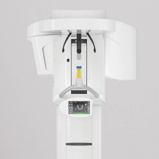
The advanced Orthophos S 3D system with conical beam.
Professionals specialised in dental imaging diagnosis.
Direct communication between the dentistry and radiology teams for coordinated care.
Whether to plan orthodontic treatment, place an implant or detect hidden cavities, dental radiography is a key tool for taking care of your oral health.
Fast track your treatment
To book an appointment or speak with one of our friendly team, please get in touch using the options below
Dental radiology is a diagnostic imaging technique that allows us to visualise the internal structures of the mouth: teeth, roots, jawbone, joints and surrounding tissues.
At Turo Park Clinics we use digital radiology, which offers multiple advantages over traditional methods:
Less exposure to X-rays (up to 90% less radiation).
Instant, high quality images.
Faster and more convenient process.
Easy storage and digital access for clinical follow-up.
Cutting-edge technology: Orthophos S 3D
Always at the forefront of innovation, at Turo Park Clinics we have the most advanced equipment on the market. Our clinic is equipped with an Orthophos S 3D cone beam, a state-of-the-art device that represents the evolution in dental imaging.
This system:
Combines the advantages of a panoramic X-ray with those of a computed tomography (3D CT).
It allows a 3D digital reconstruction of the oral structures.
It scans the entire volume in a single pass, or focuses only on a specific area, which significantly reduces radiation exposure.
It provides much more accurate images than a traditional panoramic, being especially useful in implants, complex endodontics and dental surgeries.
In our daily practice, this technology allows for more accurate diagnoses and safer treatments for our patients.
What types of dental X-rays do we perform?
Each type of X-ray has a specific purpose. In our clinic we have all the formats necessary for a complete diagnosis:
Panoramic X-ray (Orthopantomography)
General image of the whole mouth: teeth, jaws, joints and maxillary sinuses.
Ideal for detecting cysts, extensive caries, retained teeth or planning orthodontics and implants.
In this real-life case, Dr Claudia Wand demonstrates why a standard visual examination at the dentist’s is not enough. A patient came to our clinic suffering from severe chronic migraines caused by an impacted tooth that had never come through:
Why was dental radiology essential here?
Reveals the invisible: It allowed us to see through the jawbone that the tooth was impacted, twisted and pressing on delicate nerve endings.
Tackle the cause, not the symptom: Without the digital X-ray, the patient would have continued taking migraine painkillers for the rest of their life, unaware that the real root of the problem lay in their mouth.
This is the key to prevention: This case highlights the risks of skipping regular check-ups. A routine X-ray carried out in good time would have detected the abnormality years earlier, sparing the patient months of unnecessary pain and suffering.
Immediate diagnosis: Thanks to our digital technology, the patient received their X-ray results instantly during their first visit, resolving a medical mystery quickly and accurately.
When is a dental X-ray recommended?
A dental X-ray may be necessary in different clinical situations. Some of the most frequent cases are:
Detection of caries not visible to the naked eye.
Evaluation of infections or abscesses.
Study of dental development in children.
Control before and after orthodontic or implant treatments.
Examination of roots and adjacent structures in endodontic treatments.
Our answers to the most frequently asked questions about dental radiology
Are dental X-rays safe?
Las radiografías dentales exponen a los pacientes a un nivel muy bajo de radiación. Por lo tanto, son seguros para adultos y niños. Además, la tecnología de rayos X ha evolucionado considerablemente a lo largo de los años, al igual que las directrices y prácticas de seguridad.
Can I have a dental X-ray if I'm pregnant?
In most cases they are usually avoided during pregnancy. If it is urgent, specific protective measures will be taken.
How long does it take to get the results of a dental X-ray?
The result is immediate.Images are instantly available on screen for your dentist to evaluate your case and review with you.
How should I prepare for a dental X-ray?
Dental X-rays do not require any special preparation. Fasting is not necessary. You can eat and drink normally. However, if you are pregnant or suspect that you are pregnant, you should make this clear before the examination.
When should a panoramic dental X-ray be taken?
The orthopantomogram (dental panoramic) is a recommended examination in many situations. In the case of pain, for example, this examination can reveal the presence of an injury or disease. However, the orthopantomogram can also be used to examine wisdom teeth and impacted teeth in detail.
")